


Osteochondrosis of the breast is a common degenerative disease. There are specific symptoms of thoracic osteochondrosis, indicating a pathological onset. In the early stages, the patient does not worry much, so do not rush to seek help from a specialist. Over time, the symptoms worsen, forcing the patient to go to the doctor, who found a negligible pathology. You need to know what early symptoms of osteochondrosis are diagnosed and what treatment methods are most effective.
What is thoracic osteochondrosis and how it occurs
Osteochondrosis of the thoracic region is characterized by the occurrence of destructive-dystrophic processes in the middle part of the spine. The destruction is between the 8th and 19th verbs. Accurate diagnostic tests are needed to find out which vertebra is affected. Osteochondrosis of the thoracic region is often accompanied by huge complications, including prolapse or hernia. Without complications, the disease is rare, because the destruction of cartilage tissue inevitably leads to the destruction of the entire vertebral frame.
When a patient develops circulatory disorders or age-related joint wear and tear, the fibrous ring in the intervertebral disc cavity begins to collapse, losing its normal structure. Because destruction is slow, microcracks appear in the early stages where the pulposus nucleus leaks.
When the internal component leaks, the ring fibrosis begins to weaken, causing it to gradually stretch and break. When the pulposus nucleus is removed, an intervertebral hernia, the most common complication of osteochondrosis, occurs. Pathology involves damage to cartilage tissue that causes significant discomfort. Severe low back pain is also associated with neurological syndromes that develop from nerve root compression or irritation.
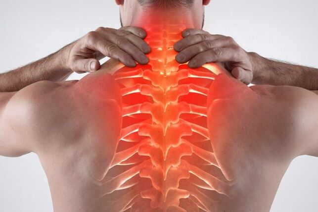
Symptoms of thoracic osteochondrosis
At the initial stage, the patient does not feel uncomfortable, so at this stage, the disease can be detected only by chance. Like other pathologies, the disease has many hidden symptoms.
Symptoms of thoracic osteochondrosis are manifested by the following manifestations:
- It's hard to breathe. Problems arise, manifested by shortness of breath and a feeling of shortness of breath. This indicates damage to the thoracic vertebrae and spinal cord.
- The main symptom is pain in the chest. There is a feeling of pressure in the heart, reminiscent of an ischemic attack.
- Anxiety occurs when the back is bent. As the disease progresses, the pain increases in this condition.
- There is a feeling of coldness in the lower or upper extremities against the background of deteriorating blood circulation.
- Pain in the chest against the background of intervertebral hernias. Anxiety is often felt more strongly on the left or right side of the affected area.
- Anxiety and swallowing problems in the throat. If there is irritation of the nerve endings in the upper part of the chest, a cough appears.
- Women may have chest pain that is not related to cyclical changes or hormonal imbalances.
- Tingling or burning sensation appears in the area of the legs and feet.
- Hair and nails become brittle and pale.
- Herpes zoster is less common.
- Back and chest pain occur at the same time.
- Less commonly, there is discomfort in the stomach, liver or pancreas.
- The onset of severe pain in the ribs, which indicates intercostal neuralgia.
- There are signs of thoracic chondrosis and compression - a similar pathology.
- There are problems with the gastrointestinal tract. Feels nausea and heaviness in the stomach.
- Men may lose several libido. There are problems in the genitourinary tract.
- Serious discomfort occurs when standing or sitting for long periods of time.
- There is a severe headache accompanied by dizziness. Aura migraines may appear.
- The patient often develops intercostal neuralgia.
- The pain may radiate to the neck or back.
If you find general thoracic osteochondrosis and its symptoms or some of them, you should immediately consult a therapist, neurologist, orthopedist. Also, such symptoms should be alerted if there are no problems in the gastrointestinal tract, cardiovascular system and lungs.
Acute and subacute symptoms are also present. With exacerbation of osteochondrosis of the thoracic region, if the patient experiences severe pain that deprives the patient of the ability to work and can only observe bed rest, subacute gait is slow and does not significantly limit the patient's motor activity.
An obvious sign of a slow lesion - no sharp pain. In the subacute stage, the symptoms disappear. There is no discomfort in basic body movements, including breathing, sneezing, or turning. A person does not suffer from pain during sleep, so the process of falling asleep is facilitated.
Important rules must be followed to prevent deterioration and remission of the subacute course of the disease:
- It is forbidden to lift weights.
- You can't bend sharply.
- It is forbidden to sit or stand for a long time. In this situation, an unconscious person often takes a position that is harmful to the spine, so there is an excessive load on the chain that causes another inflammation.
- Avoid hypothermia. Failure to maintain a comfortable temperature for the body has been shown to exacerbate the inflammatory process. Moisture is also harmful to the joints.
The duration of the subacute course is individual. If you follow the medical recommendations, the patient will be completely free of anxiety in 2-3 weeks. If conservative treatment and rest do not help, and the patient begins to suffer from nausea, dizziness and weakness, it is necessary to immediately consult a specialist. Such symptoms indicate re-inflammation.
Degrees of development of osteochondrosis of the thoracic region
There are 4 clinical stages in the development of pathological symptoms in the early stages of the disease:
- There are no clinical symptoms at the initial stage. The first stage occurs against the background of the appearance of destructive processes in cartilage and bone tissue. In the first stage, there is no rupture or elongation of the fibrous ring, so there is no hernia. They may detect signs of primary protrusion and cartilage degeneration.
- The second stage causes minor pain or discomfort. A careful patient is looking for a doctor, so osteochondrosis of the chest area is immediately detected. People who do not want to see a specialist can still tolerate the second stage using existing treatment, but self-medication will not be enough for a long time. The most common neurological symptoms at this stage may be headache, burning in the intercapular zone, neck pain, and increased blood pressure. Also at this stage, there is an increase in degenerative destruction of the spine: a fibrous ring emerges, which leads to the emergence of intervertebral hernia of the thoracic region.
- The third stage is already difficult for the patient. Persistent neurological syndromes develop, including persistent radiation pain in the shoulder blades, arms, clavicle, and lower back. The patient may show somatic and autonomic disorders, which indicate a disturbance in the functioning of the nervous system. The patient often suffers from nausea, constant headaches, dizziness, back pain. Hidden cardiac, gastroenterological, or pulmonary symptoms of the disease may also appear. At this stage there is an active demineralization of bone and cartilage tissue. There is a tendency to damage.
- The last stage is the fourth. Against the background of osteochondrosis and hernia, irreversible consequences appear - the mobility of the intervertebral discs is completely lost, and the cartilage tissue, which has a long inflammatory pathway, is replaced by osteophytes. You need an operation to remove them.
It is better to consult a doctor with the slightest symptom so as not to bring the body to a state similar to stage 3 or 4. The sooner the disease is detected and therapy is started, the sooner the patient will return to normal and learn to live with osteochondrosis. The pathological destructive process cannot be completely stopped, but it can be slowed down by leading a healthy lifestyle and exercising daily with exercise. The more a patient sees a doctor, the harder it is to stop the severe pain syndrome associated with cartilage tissue degeneration.
Risk factors and causes of the disease
There is no clear cause for the devastating changes in the spine. An important role in the development of pathology is attributed to hereditary factors. Individuals suffering from physical inactivity have been shown to have more problems with the mountain than those who engage in regular exercise. In addition, excessive physical activity can lead to cartilage destruction at an early age.
Thinning and destruction of the intervertebral discs is closely related to the load on the spine. If the muscles are not strong enough and the spine is constantly overloaded, cartilage tissue is destroyed.
What causes osteochondrosis:
- Obesity. Excess weight puts strong weight pressure on the spine. As a result, premature destruction of bone tissue occurs.
- The presence of an anomaly in the structure of bones and cartilage. Such problems are also posed during intrauterine development.
- Congenital asymmetry of intra-articular cavities in the intervertebral joints of the type of tropism anomaly contributes to the occurrence of a degenerative-dystrophic process in the spine.
- Muscle spasm in the chest, spondylosis, chronic persistent trigger points and vascular disease. These pathologies also contribute to the development of osteochondrosis of the chest.
- Prolonged exposure to vibrations in the spine in a sitting position. An example of a job is a minibus or bus driver.
- Frequent physical exertion associated with heavy lifting. Examples are working as a loader or a professional sports activity.
- Smoking and alcohol abuse. Lack of minerals and poor blood circulation in people with unhealthy lifestyles cause back problems.
- Sedentary lifestyle. Accelerated calcium leaching occurs, which is associated with insufficient physical activity and poor metabolic processes. As a result, the bones become brittle. In addition, muscle tissue atrophy due to excessive load on the spine. The result is pain, often with minimal physical exertion.
Thanks to the intervertebral discs, sufficient mobility of the chain is provided. Intervertebral discs act as shock absorbers. With the development of osteochondrosis, an accelerated demineralization process occurs, vital moisture is lost from the joints. This causes anxiety and decreased mobility in the spine.
Risk factors for thoracic osteochondrosis include:
- Advanced age. Natural degeneration occurs in the elderly, so the disease is more common after 40 years.
- Women. In girls, there are periods that help to actively leach calcium from the bones - pregnancy and menopause. Without adequate pharmacological support, spinal diseases are more likely to occur.
- Presence of hormonal diseases, endocrine diseases. If the patient has diabetes or uncompensated hypothyroidism, intervertebral disc degeneration can occur at an early age.
- Long-term immobilization. If the patient is ill and has to sleep for a long time, atrophic processes occur in the muscles, which leads to back pain.
- Previous spinal injuries. As ligaments and tendons stretch, the risk of osteochondrosis in the chest area increases.
- Presence of scoliosis. In the future, poor posture can lead to serious spinal problems, including osteochondrosis and hernias.
Diagnosis of thoracic osteochondrosis
If the patient suspects back problems, it is necessary to consult a therapist. The doctor conducts a general examination of the patient, asks about complaints, measures blood pressure. If a neurological problem is suspected, the patient is referred to a narrow specialist - a traumatologist, neurologist or orthopedist.
At a meeting with a specialist, they also ask about complaints and make an initial diagnosis of the patient. Based on the visual examination, a number of diagnostic measures are prescribed, including the following.
- Radiography. With the help of an X-ray you can assess the general condition of the skeletal system. If the patient has a hernia or osteochondrosis, the pathological indications can be taken into account - the distance between the intervertebral discs will be reduced, and sometimes there is darkening at the site of the alleged hernia. If the results of the image do not match the specialist, you should continue to look for the cause of pain and discomfort.
- CT or MRI. The most accurate diagnostic methods that allow you to accurately investigate the focal state of inflammation in the figure. A more detailed image can be seen on MRI, but if there are contraindications (the presence of a pacemaker or prosthesis in the joints), computed tomography is prescribed. CT is an advanced x-ray that allows you to see the bone, tendon and ligament in detail. The image shows the image in the form of a three-dimensional image, so the details of the damage are clearly visible.
- Biochemical and general blood test. These tests are needed to assess the patient's health. If there is an increase in leukocytes, if ESR is found, it indicates an active inflammatory process in the body. With active destruction of bone tissue, a decrease in calcium levels and a deficiency of cholecalciferol (vitamin D3) are present in the blood.
- Spinal scintigraphy. The research method detects active destruction of bone tissue. Weak bone tissue is very sensitive to fragility. The method will detect the tendency and signs of degeneration.
You should consult an experienced specialist to diagnose the disease. A complete clinical picture is needed for the final diagnosis, taking into account several laboratory research methods.
Thoracic osteochondrosis of the spine requires differentiation along with the following pathologies:
- Dishormonal spondylopathy.
- Pathologies of the urinary system, including urolithiasis, cystitis or pyelonephritis.
- Diseases of the cardiovascular system, except for sinus arrhythmia, tachycardia and angina pectoris.
- Diseases of the gastrointestinal tract, including chronic pancreatitis, gastric and duodenal ulcers, irritable bowel syndrome.
- Previous injuries, fractures.
- Tumors in the chest, including a malignant process.
- Rheumatoid arthritis (determined by C-reactive protein, rheumatic test and blood test for ESR).
- Osteomyelitis of the spine.
- Acute inflammatory process.
- Ankylosing spondylitis.
- Spondylolisthesis.
Treatment of osteochondrosis of the thoracic spine
An integrated approach to therapy is needed to slow the progression of the disease. In the early stages, only conservative therapy is indicated, consisting of the use of drugs and physiotherapy methods. In advanced cases, an operation is prescribed if the patient has a large hernia and a degree of bone degeneration. Do not self-medicate at home. Folk remedies do not eliminate thoracic lumbar osteochondrosis.
In what cases is the operation performed?
Osteochondrosis of the thoracic region adversely affects the patient's quality of life. Given the lack of effect of drug treatment on the patient, if there is a constant concern that interferes with normal life, a surgical solution to the problem may be suggested.
Absolute indicators for the operation include:
- Lack of sensitivity in the bladder and intestines.
- If there is no sensitivity in the legs and the patient loses the ability to move independently.
- Paralysis due to severe overgrowth of the hernia.
In other cases, the patient decides to remove the hernia independently. If the disease is really severe and the patient's condition does not improve with conservative treatment, doctors recommend surgery.
Drug treatment of thoracic osteochondrosis
During the exacerbation, the attending physician prescribes a variety of drugs that need to be used to eliminate the inflammatory process. The acute period is characterized by severe pain that can only be relieved with medication. If enough medication is taken, the patient recovers. Only an experienced specialist can prescribe medication; self-medication is unacceptable.
Thoracic spinal osteochondrosis is treated with the following drugs:
- Non-steroidal anti-inflammatory drugs, painkillers or analgesics. These drugs are designed to quickly relieve back pain associated with an active inflammatory process. The effect of taking a pill or injection is felt the next day. Taking any drug from the NSAID group is accompanied by side effects with long-term use, so experts recommend limiting the use of the drug for no more than 1-2 weeks. Painkillers are the most harmful to the gastric mucosa, causing gastropathy and inflammation. At-risk patients are given some medications designed to protect the gastrointestinal mucosa. Examples are proton pump inhibitors, H2 histamine receptor blockers, antacids. People with ulcers and gastritis are better off avoiding the use of NSAIDs or taking modern analogues with selective effects.
- Muscle relaxants. These drugs are very effective in the treatment of muscle spasticity. Eliminate pain associated with muscle tension. They move at trigger points located in the compressed muscle tissue. When a person is overloaded, their number increases. It relaxes the muscles well and therefore has an analgesic effect. You should take a course of medication, the average duration of therapy is at least 2-4 weeks.
- Assign B vitamins B1, B6, B12 in the form of combined injections. In large doses, these substances have a analgesic effect and have a positive effect on the nervous system. Neurotrophic drugs are effective in treating pain associated with compressed nerve roots. It is impossible to meet the norm of these substances, which are necessary to achieve a therapeutic effect with the help of nutrition, so it is prescribed in the form of drugs. The average length of a course of injections is 2-3 weeks. Then, if necessary, switch to tablets.
- Anti-inflammatory ointments, gels. If the pain is tolerable and systemic forms of NSAIDs are contraindicated, external drugs are prescribed. The advantage of external treatments is that they do not cause side effects. Rarely, skin allergies may appear, but the ointment does not cause gastrointestinal or laboratory bleeding. Another advantage of foreign products is the possibility of long-term use. You can rub on the gels for up to 4 weeks, after which they take a break. The scheme and duration of therapy is determined by the attending physician.
- Honroprotectors. These are complex substances used to nourish the cartilage tissue of the joints. The drug should be used for a long time, at least six months, after which there is a break of 2-3 months and the course of therapy is repeated. Injectable release forms are used because they are better absorbed within 2-3 months. They then undergo supportive care, including the use of tablets. It is important to understand that the drug does not stop the destruction of cartilage tissue. They create additional nutrition that slows down the degenerative processes that occur only in the bones and joints.
- Complex preparations of calcium and vitamin D3. People living in the northern latitudes have been shown not to get enough vitamin D3 because there is little sunlight in the area. To get rid of hypovitaminosis, it is necessary to take cholecalciferol supplements in winter and autumn, when solar activity is minimal. Without this vitamin, it is impossible to assimilate calcium and other minerals. Prolonged calcium deficiency causes thinning of bone tissue over time, so a person suffers from osteochondrosis and other complications. Calcium and D3 are better absorbed in combination, so complex drugs are prescribed. The dose and course of administration should be determined by the attending physician.
In addition to treatment, homeopathy, antispasmodics and complex multivitamins may be prescribed.
Conservative therapy for thoracic osteochondrosis
During the recovery period, the patient should pay sufficient attention to rehabilitation. The more carefully the patient maintains his health, the fewer attacks of the disease will occur.
The most effective conservative treatments include:
- Sports therapy. With the help of exercises, the patient learns to keep his back straight, strengthens the muscular corset. Physiotherapy can be done at any age, several times a week. The complex is selected individually, taking into account the anatomical features of the patient. Start gradually, spending no more than 5 minutes a day. As physical qualities improve, the patient learns to do more difficult exercises over a longer period of time.
- Supporting corset. Anatomical devices serve to support weakened muscles if they are contraindicated. The patient chooses a dressing depending on the neck and the type of appointment. The attending physician should select the appropriate model. The duration and form of wear is determined individually. You can't wear a corset for hours, otherwise your back muscles will weaken even more.
- Massage. In medical practice, massage is one of the most popular and effective methods of conservative treatment in the presence of osteochondrosis of the chest in a patient. Muscles need extra support during recovery. It is useful when blood flow is temporarily improved and over-stretched muscles are used with the right technique. You must attend specialist meetings several times a year during the courses.
- Physiotherapy. Physiotherapy procedures are widely used in trauma, orthopedic and neurological practice. With the help of procedures, local blood flow is improved, systemic drugs are used externally, and the apparatus affects the damaged tissues. As a result, the muscles are warmed up and the chronic inflammatory process in the affected area is eliminated. Examples of medical procedures - magnetotherapy, shock wave therapy, electrophoresis.
Less common, manual treatment and acupuncture are prescribed.
Osteochondrosis of the breast is a serious disease. It is necessary to treat the pathology thoroughly to prevent the acute progression of the disease.